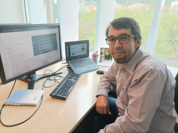
If there is one person who can make you feel the deep connection between the human body and the Earth, it is Josep de la Puente. But he isn’t an expert in the practice of grounding, a therapy that involves walking barefoot to return you to your natural state. Far from it. De la Puente (Barcelona, 1979) says that seismic waves passing through the planet and ultrasound waves passing through the human body follow the same physical principles. This helps explain how this physicist, an expert in computational seismology, ended up diagnosing breast cancer using a new technique based on ultrasound, which is harmless to women.
It all started with a conversation he once had with two friends under a tree during a trip to the Netherlands. It was at that moment, in the exchange of ideas between colleagues (Josep, Lluís and Óscar), that the possibility of developing the technology they are now using was born. His friends are also physicists—Óscar Calderón and Lluís Guasch—who at the time were at Imperial College London. That fruitful conversation led to a project that De la Puente is currently coordinating, QUSTom (Quantitative Ultrasound Stochastic Tomography), at the Barcelona Supercomputing Center (BSC). The aim of this European programme is to introduce a new medical imaging modality, based for the first time on ultrasound and supercomputing, that will complement or even replace current techniques that use X-rays, such as mammography. This technology will be completely safe for patients, as it does not use any kind of radiation, and will offer superior image quality and better monitoring of tumours, among other advantages.
The working group brings together physicists, engineers and computer scientists, who collaborate with oncologists and hospitals. Interestingly, they are using the same techniques that were previously applied to detect and study earthquakes using artificial intelligence.
Specifically, they are investigating how to obtain high-quality medical images from ultrasound data. “Instead of relying on real-time images, such as those produced by ultrasound scanners used in gynaecology,” explains De la Puente, “we are striving to obtain the highest possible image quality for a better diagnosis than the current one. In this case, it takes us a few hours to obtain the image, which does not represent a significant difference in terms of diagnostic time.”
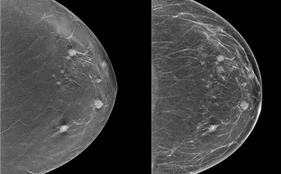
The process we use produces potentially better images than those of a mammogram, and because it is ultrasound, the test is safe for patients and very comfortable.
The procedure involves being able to simulate these sub-millimetre-sized ultrasound waves as they pass through the human body. “We repeat this process thousands of times in order to obtain images of ever higher resolution. That’s why we use the MareNostrum4 supercomputer as a computing platform,” he continues. “The process we use produces images that are potentially better than those of a mammogram, in addition to the fact that the ultrasound test is harmless for the patient and also very comfortable.”
Josep de la Puente’s professional trajectory demonstrates the importance of believing in multidisciplinarity and teamwork, as well as the value of having a way of thinking and an intelligence than can understand the relationships between very different disciplines.
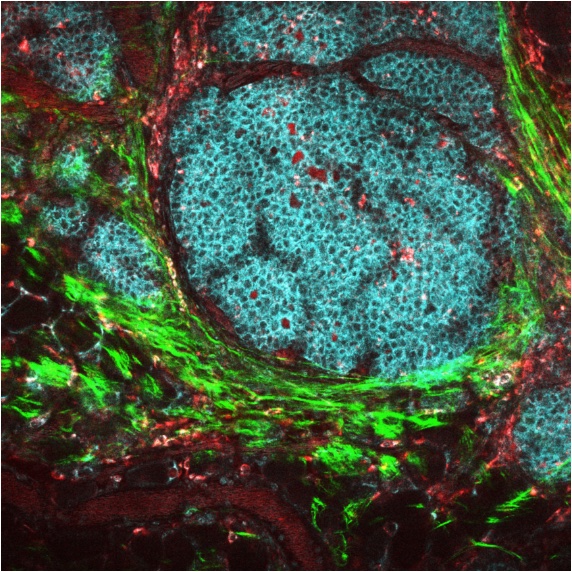
The technology being developed by De la Puente could one day help to diagnose other types of cancer and diseases. He explains how it works. “Essentially, we create a digital twin of the breast tissue and the ultrasound measuring device. This digital twin simulates any of the ultrasound emissions that are made in the physical device used by the radiologist.” And this allows them to obtain, as the scientist says, “more than just an image: it is a detailed, three-dimensional map of the tissue properties in every pixel. Wherever we look, we can obtain quantitative values of the stiffness, density or attenuation of the tissue.” By analysing these properties and the morphology or shape of the image, healthy tissue can be distinguished from cancerous tissue with a high degree of reliability.
We build a digital twin of the breast tissue, and this twin simulates any of the ultrasound emissions that are produced in the physical device used by the radiologist.
“We are currently developing the technology for breast cancer, but it wouldn’t be much different to do it for other parts of the body,” says De la Puente. “Since the images are generic, they have the potential to be used to detect other diseases that are currently the subject of medical imaging, such as bone disease or other types of cancer. Time will tell in which applications it has the most potential.”
Josep de la Puente is going to receive a machine that is the only one of its kind in the world to begin trials on this harmless technique for detecting breast cancer. What is so special about this new device? “It emits ultrasound throughout the interior of a hemispherical surface, like a basketball cut in half. When an object is placed inside, ultrasound waves are emitted from about 2,000 points along the surface. These same points also act as microphones, generating millions of acoustic signals that pass through the breast tissue, in this case, from all possible directions. This way of looking at the tissue, with omnidirectional information, is unique to this device, which was designed and built by the Karlsruhe Institute of Technology in Germany.”
But making all these innovations a reality requires a lot of people with a wide range of skills, says De la Puente. “To this end, we have several projects underway: the QUSTom project, which I coordinate at BSC and which is funded by the European Innovation Council, but at the Vall d’Hebron University Hospital, which has the capacity to validate the technology on volunteers; ARCTUR, which is helping us to reduce calculation times; Imperial College London, which proposes innovative algorithms for quality control; and, finally, Frontwave Imaging, a spin-off from BSC and Imperial College, which aims to clinically certify the technology and bring it to the market in the near future.”
There are darker aspects of this disease that should be made more visible, and one of them is the issue of dense breasts.
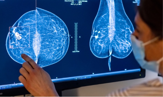
Although breast cancer has benefited from numerous awareness campaigns, there are darker aspects of this disease that should be made more visible, the expert stresses. “One of them is the problem of dense breasts. Around half of Spanish women have breasts that radiologists classify as dense. The consequence of having dense breasts is that it’s more difficult to detect incipient cancer in mammograms. In addition, and unrelated to this, dense breasts are more likely to have cancer.”
New jobs or areas of research related to this speciality are already on the horizon. “We mainly need professionals with a broad vision,” stresses De la Puente. “People who can easily relate their speciality to other fields of study. Science is becoming an increasingly collective field, where results are the fruit of collaboration. Future physicists, mathematicians, computer scientists and doctors will need to be able to communicate their ideas and receive the ideas of others in a transversal and positive way.”
We need professionals with a broad vision. Future physicists, mathematicians, computer scientists and doctors will need to be able to communicate their ideas and receive the ideas of others in a transversal and positive way.
Will new specialities emerge from these findings? “There should be training in something we could call imagology or image engineering; we need specialists in image reconstruction algorithms for all kinds of applications,” says De la Puente. “This is something that doesn’t currently exist and which I think could have an impact on medical imaging applications, but also to analyse, for example, the presence of cracks in concrete or the integrity of a complex structure such as a vehicle. It’s a very promising field of study with an important market niche, but we lack well-trained professionals who can access it.”
This expert also stresses the need to involve more women in these fields: “One thing I think is very necessary is to get more women involved in science and technology. There are more and more of them, but we have to make sure that gender is no longer an issue in this field,” he says.
Finally, we asked this researcher about his big dream, the result of so much hard work and the enthusiasm he radiates for his work: “I hope that the technologies I’m working on, both in seismology and in medical imaging, will have a real social impact. I hope to see this very soon. I also hope that I will continue to find challenges and that the team I work with will enjoy what they are doing and be proud of what they are contributing. It seems very simple, but you can’t play this game without a good team.”
Comments on this publication